Introduction
The main thrust of research performed in our laboratory is to develop new techniques for the label-free detection of biochemical events at the liquid (especially blood, serum and plasma) -solid interface. In terms of perspective the aim is to generate new technologies for application in bioanalytical chemistry and detection science, notably in the areas of clinical diagnostics, biocompatibility and nanomedicine. The work is highly interdisciplinary in character and involves activities ranging from the design and construction of new instruments to theoretical calculations. Research staff possesses multidisciplinary expertise including such areas as analytical chemistry, basic electronics, surface chemistry, biochemistry, computational methods, and materials science.
Ultra-high-frequency acoustic wave detection in bioanalytical chemistry (EMPAS)
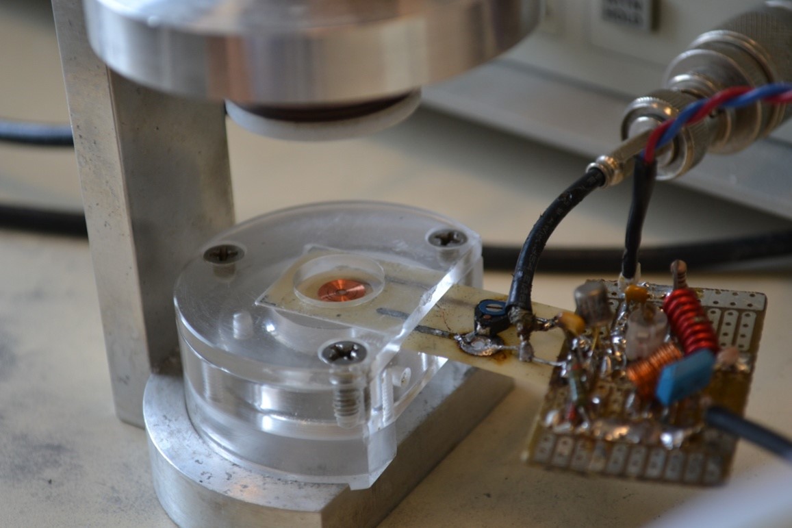
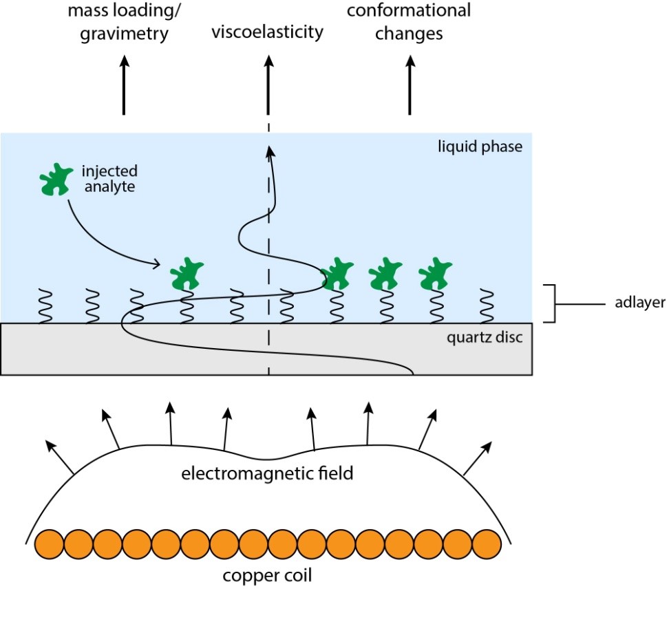
Work in this area is divided into several categories, all strongly interrelated. First, we are employing transverse shear-wave sensors to detect, in label-free format, a variety of important biochemical systems. A key feature of our detection research is the design and construction of an ultra-high frequency electromagnetic excitation configuration for application to the study of biological species at interfaces and in the field of clinical diagnostics by acoustic physics (electromagnetic piezoelectric acoustic sensor, EMPAS). Here, a specially fabricated planar spiral coil is brought close to a piezoelectric disk which, in turn, is incorporated into a flow-through stream. The coil sets up a very high frequency electromagnetic field, which couples into the piezoelectric tensor of the disk. Such an arrangement allows acoustic physics to be operated at frequencies of 1 GHz, and is, therefore, extremely sensitive from an analytical chemical standpoint. This revolutionary instrument offers huge advances in the detection of biochemical events because of the high frequency, tuneability and advantageous use of quartz as a substrate for surface chemistry. Current research also involves use of devices based on lithium niobate and the improved and enhanced design of the instrumental and electronic aspects of the technology.
Interaction of surfaces of biosensors and medical devices with biological fluids: anti-fouling and biocompatibility
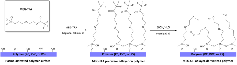
A crucial aspect of biosensor technology, which shares many common features with the field of biocompatibility, is the unwanted adsorption, or fouling, by biological species when devices are immersed in blood, serum and plasma. In our work we are both interested in methods to prevent such fouling and in novel methods for the attachment of proteins and nucleic acids to various materials with applications in both fields. This involves the synthesis of new silanes and other linker moieties with a view to not only probe binding for biosensor development but also the tandem avoidance of non-specific adsorption from entities present in biological fluids. Research with linker molecules is centered on the physical chemistry of probe-diluent ratio, probe biological activity and adsorption phenomena. Recently we have demonstrated that a 0.5 nm silane film can be successfully applied to biosensor surface in order to act in an anti-fouling capacity.
With respect to biocompatibility, we are researching the surface modification of polymeric materials used in medical bypass surgery, and renal dialysis. Micro-clots are known to form on the surfaces of materials used as conduits which are in contact with blood during medical procedures. These clots can have serious medical consequences in terms of cognitive disability. The analogous ultra-thin films employed for our biosensor work have been shown to be extremely successful in the prevention of thrombi. Experiments in this case involve the real-time measurement of aggregation of platelets from human blood using confocal fluorescence microscopy. The results of this research are being extended to the surface modification of stainless-steel stents. Stents are expandable cylindrical scaffolds that are implanted within narrowed arteries. They are typically constructed from medical grade stainless steel and can be implanted as bare metal stents or as drug eluting stents depending on patient candidacy. The goal of our research is to develop a biologically active and biocompatible, fully sterilizable coating for stainless steel stents that will reduce both neointimal hyperplasia and the physiological attack of the stent.
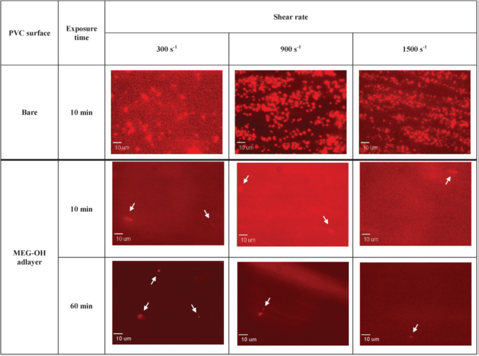
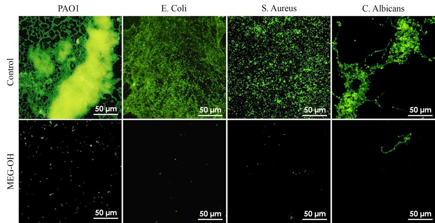
The group is also researching the application of our patented surface modification for the prevention of adhesion of bacterial microbes to polymeric materials conventionally employed for the fabrication of catheters. Indwelling urinary catheters are employed in both hospital and nursing home settings to relieve urinary retention. By far the most serious issue with regard to use of these devices is the development of nosocomial urinary tract infections (UTIs), generally known as catheter-associated UTIs (CAUTIs). These infections have been shown to occur with 80% of patients resulting not only in the subsequent severe medical sequelae, but also significant economic cost. CAUTIs can be caused by several microbial species and are usually associated with biofilms on the device produced by species such Escherichia coli, Proteus mirabilis, Pseudomonas aeruginosa, Candida albicans, and Staphylococcus aureus. Our research has demonstrated in proof-of-principle experiments that adhesion of bacteria to modified polymeric materials can be very significantly reduced. The overall goal is work towards commercialization of the technology.
Early-stage detection of ovarian cancer
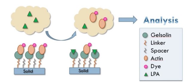
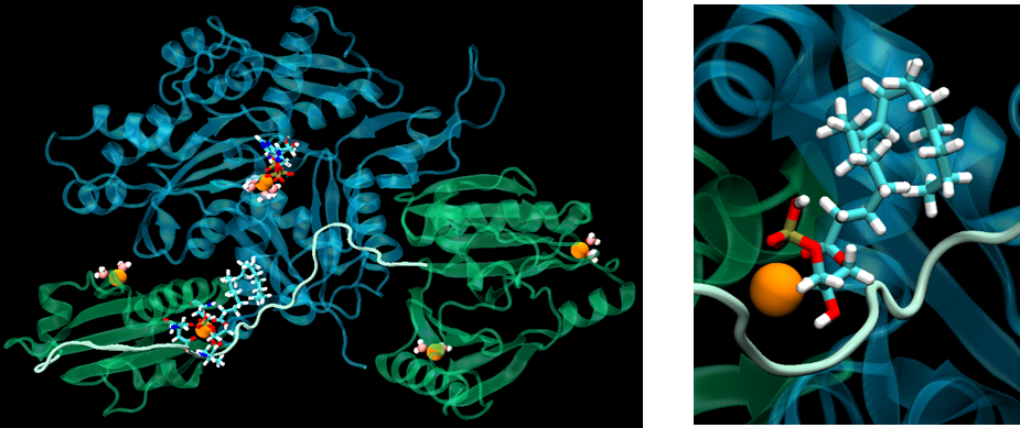
Ovarian cancer is associated with tumors that form in the tissue of the ovary, a component of the female reproductory system. Ovarian cancers are either ovarian epithelial carcinomas (cancer that begins in the cells on the surface of the ovary) or malignant germ cell tumors (cancer that begins in egg cells). Such cells can shed or spread to other organs via the process generally understood to be the mechanism of metastasis. It is anticipated to kill 14,000 women in the US in 2015 with over 20,000 new cases being diagnosed. It is often referred to as the “silent” killer since early symptoms are not radically different from certain normal conditions. The cause of the disease is unknown and is difficult to detect, with the existing CA125 assay widely considered to be inadequate. Accordingly, there is an urgent need for an analytical test that is both sensitive and selective and can be incorporated at low cost into a high-throughput instrumental system to be available in the clinical diagnostic laboratory. Our research involves the use promising biomarkers that we propose to detect via a biosensor format, such as lysophosphatidic acid (LPA). The molecule has been shown to be present in the blood of patents at all stages of the disease. LPA levels have been found to be elevated in 90% of stage I ovarian cancer patients (in the several micro-molar range), and 100% of later stage patients. We have initiated research on LPA detection using gelsolin-actin chemistry. Using fluorescence spectroscopy, we have demonstrated that gelsolin, a large protein of 782 amino acids and 6 domains, binds LPA with a Kd value of 6 nM with only the first 3 domains of gelsolin being necessary for both actin and LPA binding, thus reducing the size and complexity of the probe. This binding capability is ideal for detection by a simple spectrofluorometric assay and/or by biosensor. We are imposing the protein-actin complex on the biosensor with a view to monitoring the removal of actin via LPA binding from LPA spiked serum.
We are also looking at transfer of the spectroscopic approach to detection to one based on electrochemical biosensor technology. Different techniques using the affinity-based approach to detect the using aforementioned gelsolin-actin chemistry are being employed. We are following two different strategies: a) a label-free approach using electrochemical impedance spectroscopy (EIS), and b) a redox-labeling approach using square wave voltammetry (SWV). The main challenge in this work is achieving the limit of quantification (LOQ) as low as 1 µM.
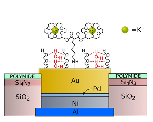
Microelectrode Measurement of K+ and H+ in Nerve and Brain Tissue
Abnormal values of cations such as K+ and H+ in tissue have been associated with several conditions such as epilepsy, neuromuscular ataxia, cardiac arrhythmia, and Alzheimer’s disease. This observation, in addition to fundamental studies in neuroscience, has resulted in attempts to monitor the in vivo concentration of cations. Such measurements enable temporal and spatial resolution of neural activity, including the peripheral nervous system. In addition, the technique offers the possibility to chronically record cation concentration as an implantable sensor in order to function in a closed-loop fashion for organ monitoring and control. Our research is composed of 2 related strategies – measurement of cations in nerves (in collaboration with the University of West London, UK) and brain tissue (in collaboration with the Krembil Research Institute, Toronto). With respect to the former, the future application is with respect to assay of the 2 cations in the vagus nerve. This nerve, which is the longest in the nervous system, controls many functions, for example satiety, via the brain stem. The second area of interest is concerned with the role that pH and potassium play in epilepsy. The reason for this is, for example, that the extracellular concentration of K+ changes during seizures so its real time assay is of utmost importance. In our research we are using both microelectrodes constructed from planar and wire-based metals such Gold and Iridium. In both cases a proprietary probe for a cation such K+ is surface-attached to the electrode in tandem with an antifouling agent such as described above. These microelectrodes can be implanted in tissue for real time measurement of the cations. Future research will involve use of the electrodes in a closed loop configuration.
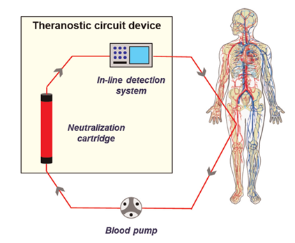
Theranostics: detection and removal of endotoxin from biological fluid
For many years the traditional approach in medicine has been to diagnose a disease condition which is often followed by drug and other treatments in order to ameliorate the factors that cause such a condition. However, many of the troubling diseases of our time are highly heterogeneous in their expression, which results in the fact that a number of drug-based therapies are only effective for certain sub-populations of the afflicted. These factors have resulted in the appearance of modern-day “personalized medicine” and one rapidly developing aspect of this approach is theranostic technology, that is, the application of therapy in intimate connection with clinical diagnostics. In our work, we are producing an off-line biosensor assay of lipopolysaccharide (endotoxin or LPS) for coupling with a cartridge which is designed to remove this molecule from patient blood via hemoperfusion. LPS is responsible for the medical condition of endotoxemia or sepsis and is a major trigger of shock, possibly leading to death. The biosensor is based on the afore-mentioned EMPAS system which incorporates a polymyxin surface-attached probe for operation in tandem with our proprietary anti-fouling chemistry. We are also working on a completely new cartridge for the on-line removal of LPS which involves the use of micro-bead technology and immobilized polymyxin.